Fig. 1.
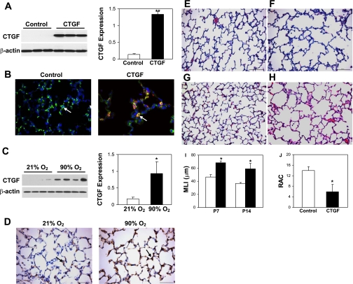
Connective tissue growth factor (CTGF) disrupted alveolarization. Western blot analysis demonstrated a significant increase in CTGF expression in CTGF- (A) and hyperoxia-exposed lungs (C). B: double immunofluorescence staining was performed on lung tissue sections at P14 with an anti-pro-surfactant protein C (SP-C) (green signal) and an anti-CTGF antibody (red signal) and 4,6-diamidino-2-phenylindole (DAPI) nuclear staining (blue signal). Pro-SP-C (arrow) was detected in control and CTGF lungs, but CTGF was only colocalized with pro-SP-C (orange signal, arrow) in CTGF lungs. D: immunostaining with an anti-CTGF antibody demonstrated a marked increase in CTGF expression in hyperoxia-exposed lungs compared with normoxia-exposed lungs. Hematoxylin and eosin (HE)-stained lung tissue sections from control lungs at postnatal day (P) 7 (E) and P14 (G) demonstrated normal alveolar development. However, overexpression of CTGF disrupted alveolar development with larger and simplified alveoli at P7 (F) and P14 (H). Mean linear intercept (MLI) was significantly increased in CTGF lungs at P7 and P14 (I). Open bars, control lungs; solid bars, CTGF lungs. Radial alveolar count (RAC) was significantly decreased in CTGF lungs (J). N = 4/group. *P < 0.01. **P < 0.001. Magnification: ×40 (B, D), ×20 (E–H). Scale bars: 50 μm (D, G, H) and 100 μm (E, F).

