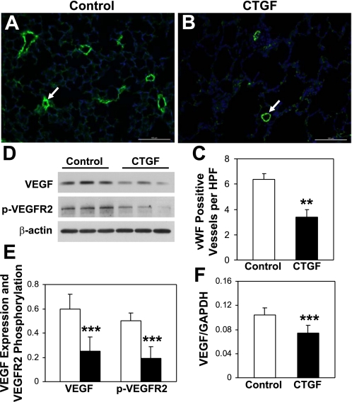
Fig. 2.
CTGF decreased vascular development. Immunofluorescence staining was performed on lung tissue sections from control (A) and CTGF (B) lungs at P14 with an anti-von Willebrand factor (vWF) antibody (green signal) and DAPI nuclear staining (blue signal). The number of vWF-positive vessels (15–50 μm, arrow) was significantly decreased in CTGF lungs (C). Western blot analysis (D and E) demonstrated significant decreases in VEGF expression and VEGF receptor 2 (VEGFR2) phosphorylation in CTGF lungs (solid bars) compared with control lungs (open bars). Real-time RT-PCR demonstrated a significant decrease in VEGF gene expression in CTGF lungs (F). N = 4/group in C and 3/group in E and F. **P < 0.001 and ***P < 0.05. Magnification: ×20. Scale bars: 100 μm.

