Fig. 3.
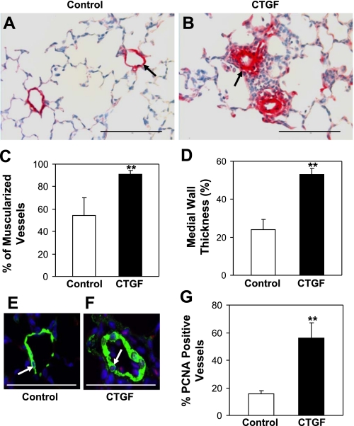
CTGF induced pulmonary vascular remodeling. Immunostaining with an anti-α-SMA antibody was performed on control (A) and CTGF (B) lungs at P14. Overexpression of CTGF significantly increased the percentage of muscularized vessels (C) and medial wall thickness (D) (15–50 μm). Double immunofluorescence staining was performed with an anti-α-smooth muscle actin (α-SMA) (green signal) and an anti-proliferating cell nuclear antigen (PCNA) antibody (red signal) and DAPI nuclear staining (blue signal). E: a representative small pulmonary vessel from control. F: representative small pulmonary vessel from CTGF lung. Quantification of PCNA-positive vessels (pink signal in nuclei, arrow) was significantly increased in CTGF lungs (G). N = 4/group. **P < 0.001. Magnification: ×40. Scale bars: 100 μm (A and B); 50 μm (E and F).

