Fig. 6.
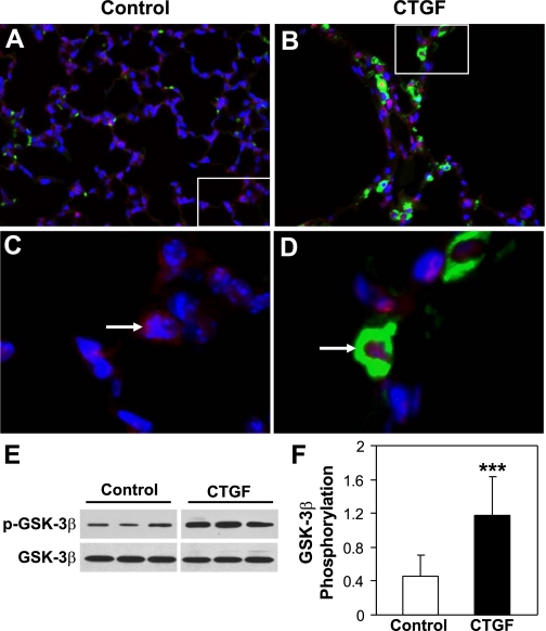
CTGF induced β-catenin nuclear translocation and GSK-3β phosphorylation in vivo. Double immunofluorescence staining for β-catenin (red signal) and CTGF (green signal) as well as DAPI nuclear staining (blue signal) was performed on lung tissue sections from 2-wk-old control (A and C) and CTGF (B and D) lungs. In control lungs β-catenin was mainly detected in the cytoplasm, whereas in CTGF lungs β-catenin was largely detected in the nuclei (pink signal) and that is colocalized with CTGF in the cytoplasm. Western blot analysis demonstrated that overexpression of CTGF significantly increased Ser9 GSK-3β phosphorylation (E and F). N = 5/group, ***P < 0.05. Magnification: ×40 (A and B). C: focal enlargement of A. D: focal enlargement of B as indicated with same magnification.

