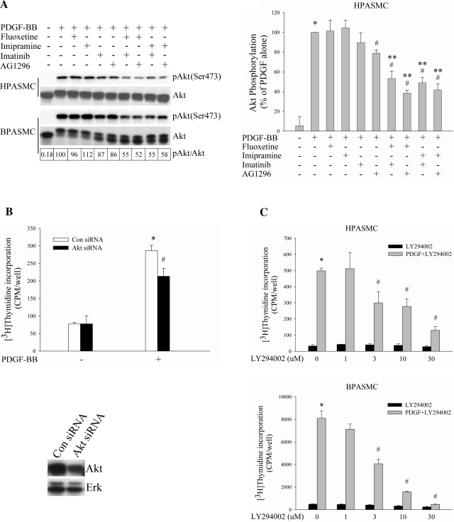
Fig. 3.
SERT participates in PI3K/Akt pathway in PDGF-stimulated PASMC proliferation. A: SERT inhibitors augment the blockade of PDGF-induced Akt phosphorylation by PDGFR inhibitors alone. Serum-starved HPASMCs and BPASMCs were preincubated with 10 μM fluoxetine, 10 μM imipramine, 1 μM imatinib, and 10 μM AG1296 individually or in combinations for 30–60 min depending on inhibitors and then were treated with 10 ng/ml PDGF for 10 min. Phosphorylation of Akt was determined by Western blot analysis of the whole cell lysate using phospho-specific antibodies, quantified as the ratios of phospho-Akt and Akt, and expressed as percentage of PDGF treatment alone (as noted for BPASMCs in boxes at bottom of figure). Bar graphs represent means ± SD for 3 independent experiments in HPASMCs. B: inhibitory effect of Akt siRNA on PDGF-induced HPASMC proliferation. Akt and Erk expressions were detected by Western blotting using Akt and Erk antibody, respectively. Bar graphs shown are means ± SD, n = 4. *Significant difference from cells without PDGF treatment (P < 0.05). #Significant difference from Con siRNA cells plus PDGF treatment (P < 0.05). C: PI3K inhibitor LY294002 dose dependently blocks HPASMC and BPASMC proliferation by PDGF. Quiescent cells were incubated with LY294002 for 60 min at indicated concentrations and then treated with 10 ng/ml PDGF for 24 h. Shown are means ± SD for n = 3. *Significant difference from untreated cells (P < 0.05). #Significant difference from cells treated with PDGF alone at P < 0.05. **Significant difference from cells treated with inhibitor alone plus PDGF (10 μM fluoxetine, 10 μM imipramine, 1 μM imatinib, and 10 μM AG1296 plus 10 ng/ml PDGF, respectively) at P < 0.05.

