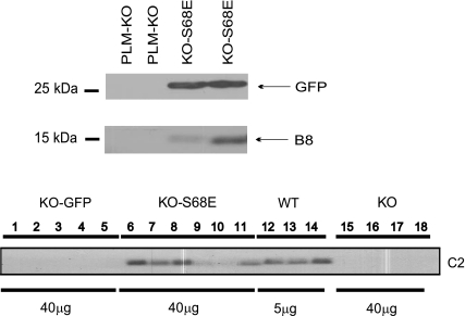
Fig. 2.
rAAV9-mediated expression of GFP and PLM S68E mutant in PLM-KO hearts. Top: 5 wk after rAAV9-S68E or saline injection, LV homogenates were prepared and subjected to SDS-PAGE followed by Western blot analysis. GFP and PLM S68E mutant (detected with B8 antibody) were present in rAAV9-S68E- but not saline-injected PLM-KO hearts. Bottom: PLM or PLM S68E mutant (identified with C2 antibody) was present in wild-type (WT) and KO-S68E LV homogenates, respectively, while no C2 signal was detected in KO-GFP or PLM-KO (injected with saline) hearts. Note the differences in amount of protein loaded for WT (5 μg) and KO (40 μg) LV homogenates. Note also that WT PLM and PLM S68E mutant have similar molecular masses since expressions of GFP and PLM S68E mutant were driven by 2 separate promoters.

