Fig. 2.
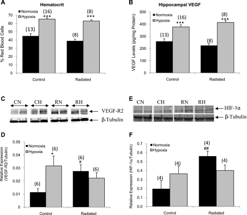
Confirmation of hypoxic status of animals. Hematocrit (A) and vascular endothelial growth factor (VEGF, B) protein levels increased in response to hypoxia treatment in both groups (n = 8–16 animals/group). C: representative Western blot for VEGF receptor (R) 2 levels. D: quantification of Western blot analysis showed that VEGF-R2 protein levels increased in the control animals in response to hypoxia and also increased in response to radiation (n = 6/group). E: representative Western blot for cortical hypoxia-inducible factor (HIF)-1α in nuclear fractions. F: quantification of the Western blot showed that, in the normoxic-treated radiated group, nuclear HIF-1α is increased compared with controls (n = 4/group). CN, control normoxia; CH, control hypoxia; RN, radiated normoxia; RH, radiated hypoxia. *P < 0.05 and ***P < 0.001 vs. the normoxic group. #P < 0.05 and ##P < 0.01 vs. the control normoxic group.

