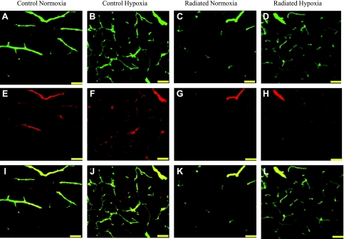
Fig. 3.
Representative images of CD31 and smooth muscle actin (SMA) staining in blood vessels within the hippocampus. Projection image showing SMA (A–D), CD31 (E–H), and merged staining of vessels within CA3 of the hippocampus (I–L) (scale bar = 50 μm). Images were captured using confocal microscopy at ×40 magnification. Analysis revealed reduced CD31 and SMA capillary density in subregions of the hippocampus in response to radiation and complete recovery of CD31 and SMA density in response to hypoxia treatment.

