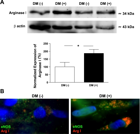
Fig. 3.
A, top: Western blot analysis of the expression of arginase 1 (Arg 1) in coronary arterioles isolated from the atrial appendage of patients with (n = 5) and without (n = 5) DM. Anti-β-actin was used to normalize for loading variations. A, bottom: summary of normalized densitometric ratios. Data are means ± SE. B: representative images of individual endothelial cells (×100, 1.4 numerical aperature objective) on acetone-fixed, en face coronary arteries obtained from patients without (DM−, n = 4; left) or with (DM+, n = 4; right) DM. Cells were immunolabeled with primary antibodies against endothelial nitric oxide synthase (eNOS) and Arg 1. Fluorescent labeling of anti-eNOS was performed with Alexa 488 secondary antibody (shown in green) and Alexa 597 antibody (shown in red) for anti-Arg 1. 4,6-Diamidino-2-phenylindole (DAPI) was used for nuclear staining (shown in blue). Coimmunostaining is shown in orange/yellow in the merged images; cells represent 5 cells from 3 different regions in each patient.

