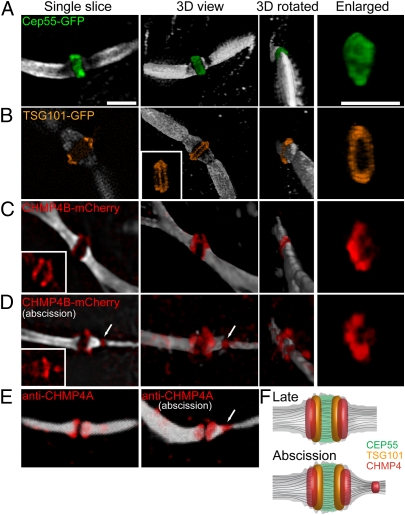
Fig. 2.
Spatial organization of the ESCRT complex in the midbody determined by SIM. MDCK cells expressing CEP55-GFP (A), TSG101-GFP (B), or CHMP4B-mCherry (C and D) were synchronized, fixed, stained with anti–α-tubulin antibodies, and imaged by SIM. (A–D) Each panel shows (from left to right) a single slice, a 3D rendering, a 3D rendering rotated 90°, and a zoomed-in image of the structure. Microtubules are colored in white, CEP55-GFP in green, TSG101-GFP in orange, and CHMP4B-mCherry in red. (A) CEP55 form a diffusely filled structure that is 1.4 ± 0.15 μm in diameter and 0.75 ± 0.07 μm in width. n = 10. A similar structure was observed for MKLP1 (Fig. S3A). (Scale bar: 2 μm.) (B) TSG101 forms a tightly packed double-ring structure surrounding the microtubules at the center of the midbody dark zone (width = 0.82 ± 0.03 μm). The rings are 1.7 ± 0.07 μm in their outer diameter and are 0.23 ± 0.02 μm apart. (Inset) A rotated image demonstrating the existence of two separate rings (n = 5). (C and D) CHMP4B concentrates in two broken rings that are 0.43 ± 0.08 μm apart. The diameter of each broken ring is 1.25 ± 0.18 μm. In some cells CHMP4B also shows an additional pool that is located asymmetrically 1.2 μm away from the center of the dark zone (arrow in D) and is perfectly colocalized to the site of microtubule constriction. (Insets) CHMP4B signal alone (n = 14). The average diameter of the microtubules in all the measurements (excluding D) is 0.98 ± 0.12 μm. The larger diameter measured for the proteins localized to the dark zone correlates with the diameter measured for this area using a membrane marker (1.6 μm; Fig. S3). The dark zone (the zone of no microtubule staining) is 0.7 ± 0.1 μm in width. (E) Confocal 3D-rendered images of antibody staining of endogenous CHMP4A (red) and tubulin (white) on the intercellular bridge of dividing MDCK cells. Images are consistent with the CHMP4B-mCherry localization described by SIM (D). (F) A model for ESCRT organization at the midbody integrating the SIM measurements indicated above.

