Fig. 2.
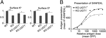
MHC class I antigen presentation is impaired in the absence of UGT1. (A) WT, KO, KO.UGT1+, and KO.UGT1− cells were stained for surface Kb/β2m (Y3) (Left) and Db/β2m (B22) (Right) for FACS analysis. Data shown are mean ± SD of two or three experiments. *P < 0.05, **P < 0.01, ***P < 0.001. (B) Presentation of SIINFEKL by Kb is impaired in the absence of UGT1. Twenty-four hours after the construct encoding GFP-ubiquitin-SIINFEKL was transfected into KO.UGT1+ and KO.UGT1−, cells were stained for Kb/β2m/SIINFEKL trimer (25-D1.16) for FACS analysis. Cells were split into eight groups according to the extent of their GFP levels. For each group, the mean 25-D1.16 levels (surface presentation of SIINFEKL) were plotted against the mean GFP levels (the relative antigen dose).

