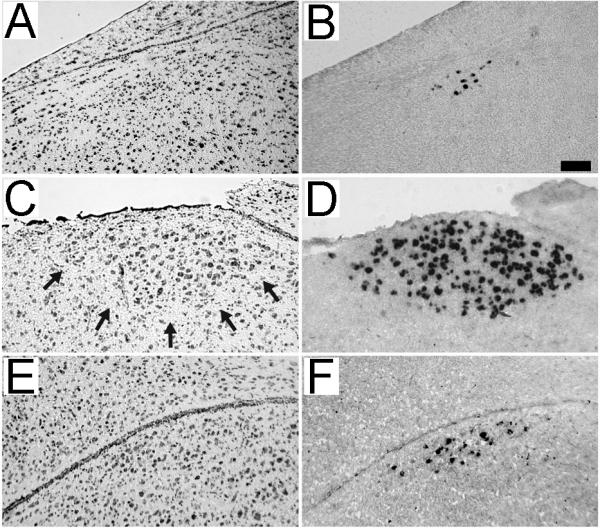
Figure 5.

Comparison of Nissl staining and zRalDH labeling. Shown are adjacent parasagittal brain sections from an adult female at the level of HVC (A and B; ~2 mm lat) and from a juvenile (20 dph) male at the level of HVC (C and D; ~2 mm lat) and paraHVC (E and F; ~0.9 mm lat). Sections were stained for Nissl (A, C and E) or processed for zRalDH in situ hybridization (B, D and F). Arrows in panel C denote the ventral boundary of HVC that is characteristic of Nissl-stained sections of male brains. In contrast, HVC in females (panel A) and paraHVC in juvenile males (panel E) are not readily visible under Nissl. Scale bar = 0.025 mm.
