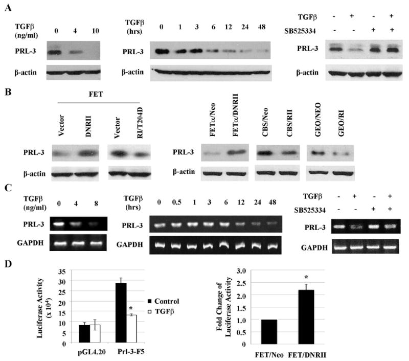
Figure 1. TGFβ signaling inhibits expression of PRL-3 in colon cancer cells.

A& C, PRL-3 protein (A) and mRNA (C) expression in FET cells treated with TGFβ for 48 or 24 hrs (left panel), 4 ng/ml TGFβ1 for the indicated time periods (middle panel) or 4 ng/ml TGFβ with or without 200 nM SB525334 for 48 or 24 hrs (right panel). Quantitation of PRL-3 from western blots and RT-PCR analyses is presented in Supplemental Figures S2A, S2B, S3A and S3B. B, PRL-3 expression in FET cells transfected with a DNRII or a constitutively active RI (RI/T204D, left panel) and in FETα, CBS and GEO cells transfected with DNRII, RII or RI respectively (right panel). Quantitation of PRL-3 from western blots analyses is presented in Supplemental Figure S2C. D, PRL-3 Promoter activity in FET cells treated with 4 ng/ml of TGFβ1 for 24 hr (left panel) and in FET Neo and FET DNRII cells (right panel). Firefly luciferase values were normalized to β-galactosidase activity. The data are presented as the mean ± SD of triplicate experiments. *P < 0.03.
