
Figure 3. PRL-3 is a direct target of TGFβ/Smad signaling.
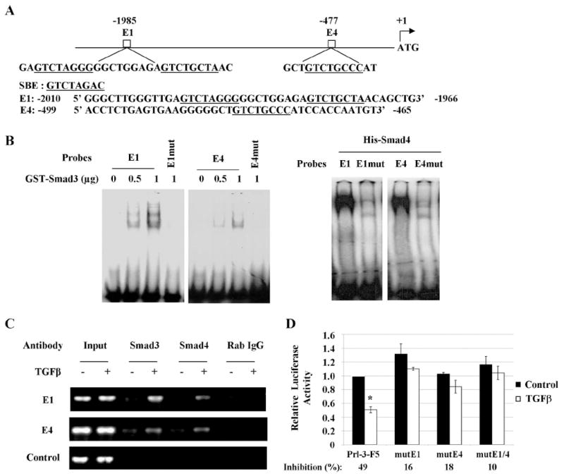
A, Schematic representation of two potential Smad binding sites, E1 and E4, in the PRL-3 promoter. SBE or SBE-like sites are underlined. B, EMSA assays were performed with oligonucleotides containing wild type or mutant E1 or E4 and recombinant proteins GST-Smad3 or His-Smad4. C, CHIP assays were performed as described in Materials and Methods. Rabbit (Rab) IgG was used as a negative control. Input represents 1% of total chromatin used in the assay. D, Relative activity of PRL-3 promoter or promoters containing mutations in E1 and/or E4. Inhibition of promoter activity by TGFβ was calculated as percentage of suppression relative to the control. The data are presented as the mean ± SD of triplicate experiments. *P < 0.02.