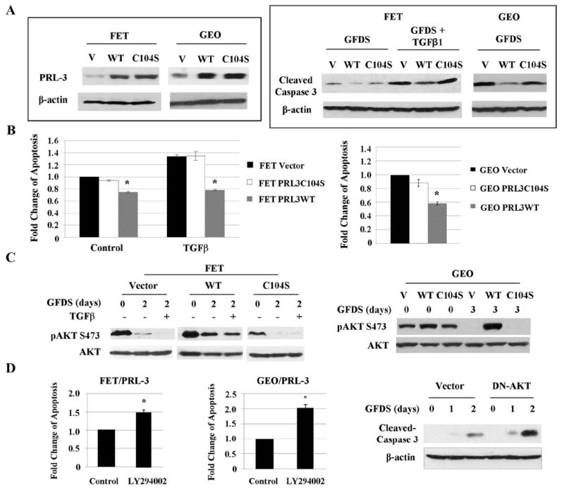
Figure 4. PRL-3 promotes cell survival under GFDS through activating the PI3K/AKT pathway.

Wild type or catalytic inactive mutant of PRL-3 was stably transduced into FET and GEO cells. A, PRL-3 expression in above-mentioned cells (left panel) and levels of cleaved caspase 3 in FET cells under GFDS with TGFβ for 2 days and in GEO cells under GFDS for 3 days (right panel). Quantitation of cleaved caspase 3 from western blots analyses is presented in Supplemental Figures S5A and S5B. B, DNA fragmentation assays in FET and GEO cells subjected to GFDS as described above. The data are presented as the mean ± SD of triplicate experiments. *P < 0.02. C, AKT expression and phosphorylation in FET cells under GFDS with TGFβ for 2 days (left panel) and GEO cells under GFDS for 3 days (right panel). Quantitation of AKT phosphorylation from western blots analyses is presented in Supplemental Figures S7A and S7B. D, DNA fragmentation assays in FET/PRL-3 and GEO/PRL-3 cells treated with LY294002 (25 nM) for 2 or 3 days respectively. The data are presented as the mean ± SD of triplicate experiments. *P < 0.01 (left and middle panels). Levels of cleaved caspase 3 were determined in FET/PRL-3 cells transfected with a DN-AKT (right panel). Quantitation of cleaved caspase 3 from western blots analyses is presented in Supplemental Figure S5C.
