
Figure 6. PRL-3 mediates metastasis of colon cancer cells in an orthotopic model.
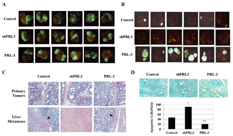
A, GFP images of primary tumors. B, GFP images of liver metastasis. C, H&E stain of primary tumors and liver metastases. Arrows indicate liver metastases. D, TUNEL stain of primary tumors. TUNEL images were captured at 10x magnification. The panels are representatives of multiple fields of tumor sections from at least 6 tumors per group. Numbers of apoptotic cells were determined as described in Materials and Methods. The data are presented as the mean ± SD. *P < 0.001; **P < 0.005.