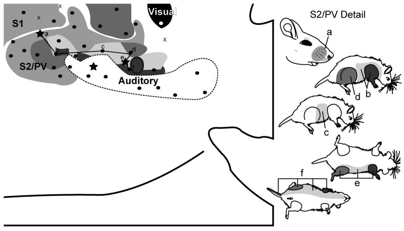
Figure 6.
Topography of the grasshopper mouse neocortex with mapping of S2/PV. The schematic of the microelectrode-derived map of cutaneous inputs to the neocortex shows a representative receptive field sequence (see supplementary Fig. 4 for complete mapping schematic). Rostral is left, medial is up for the cortical schematic.

