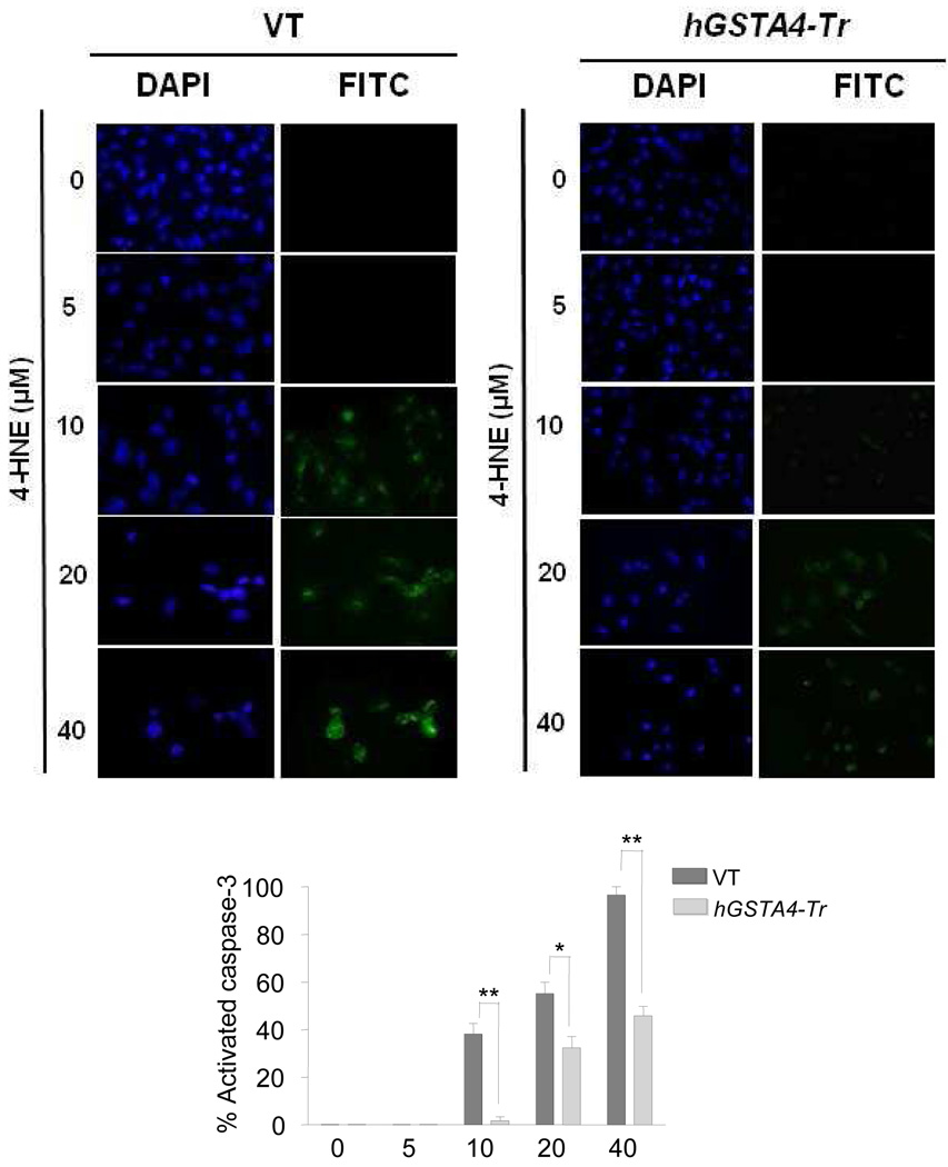
Figure 2. In situ analysis of 4-HNE mediated activation of caspase-3 in VT and hGSTA4-Tr RPE cells.
RPE cells (1 × 10⁵) were treated with 0–40 µM 4-HNE for 12 h. The activation of caspase-3 in these cells was examined by staining with 10 µM CaspACE™ FITC-VAD-FMK in situ marker according to the manufacturer’s instructions. The slides were mounted with Vectashield DAPI mounting medium and observed under a fluorescence microscope (Olympus) using the standard filter sets for DAPI and FITC. Percentage of caspase-3 activated cells presented in bar graph was determined as described previously (Duan et al., 2003). The data represent the mean ± SD (n =3).

