Figure 2.
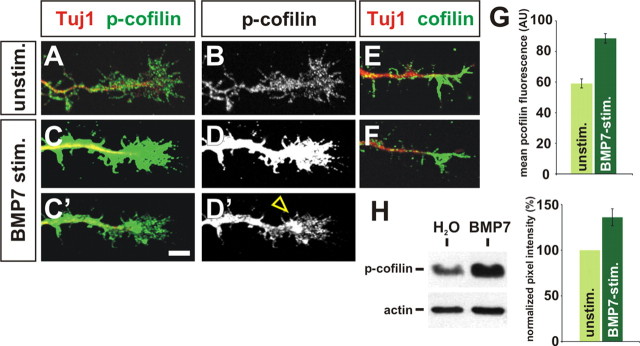
Cofilin is acutely phosphorylated after stimulation with BMP7. A–H, Cultures of dissociated E13 rat commissural neurons were briefly stimulated with either water or BMP7 recombinant protein and then labeled with antibodies against p-cofilin (green, A–D), total cofilin (green, E, F), and neuronal class III β-tubulin (Tuj1; A, C, E, F) to reveal neuronal processes (Geisert and Frankfurter, 1989) or subjected to a Western blot analysis (H). A, B, In unstimulated culture conditions, p-cofilin is evenly distributed at low levels across commissural growth cones. C, D, In contrast, the level of p-cofilin was dramatically increased in commissural growth cones treated with BMP7 (open arrowhead), suggesting that the inactivation of cofilin in commissural growth cones is an immediate consequence of BMP7 treatment. A–D were imaged using identical settings on the confocal microscope, whereas C′ and D′ were imaged at a lower gain/offset levels to reveal the distribution of p-cofilin in the commissural growth cone after stimulation. E, F, The distribution and levels of total cofilin protein is similar in unstimulated (E) and BMP7-treated (F) commissural growth cones, suggesting that BMP7 treatment directly results in the phosphorylation of cofilin rather than a redistribution of cofilin protein. G, Quantification of mean p-cofilin immunofluorescence in stimulated and unstimulated commissural growth cones. There was a significant increase (p < 2.1 × 10⁻⁹, Student's t test) in the level of p-cofilin after addition of BMP7 (n = 162 neurons) compared with unstimulated control cultures (n = 117 neurons). AU, Arbitrary units. H, Western blot of dissociated E13 neurons stimulated with either water or BMP7. There was significant increase (p < 0.0023) in the level of p-cofilin after BMP stimulation (n = 4 experiments). Scale bar: A–F, 10 μm.

