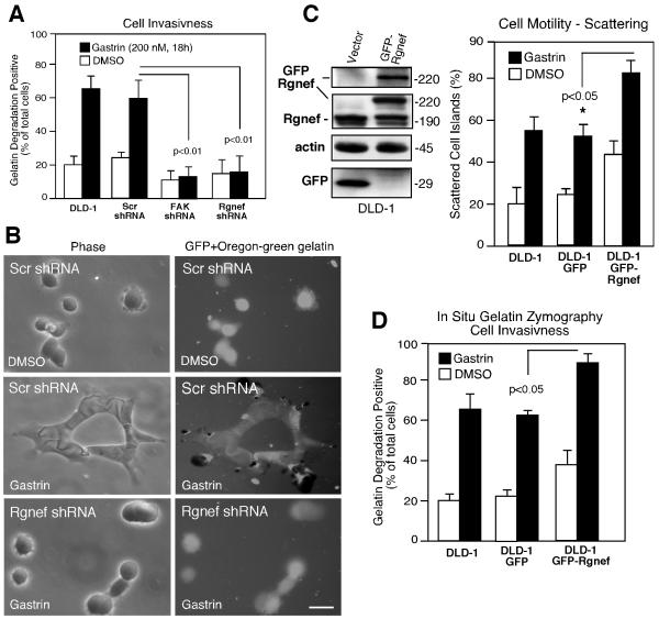
Figure 4.
Rgnef and FAK facilitate gastrin-stimulated DLD-1 matrix degradation. A, In situ gelatin zymography analyses after DMSO (control) or gastrin addition were performed with parental DLD-1 cells or the indicated shRNA-expressing DLD-1 cells and analyzed by microscopy. Values represent percent of cells with associated gelatin degradation patches. Values are the mean +/− SD from 3 experiments. Statistical significance is compared to Scr shRNA DLD-1 cell results. B, Representative phase and fluorescent images of Scr- or Rgnef shRNA-expressing DLD-1 cells plated on Oregon green gelatin under DMSO (control) or gastrin-treated conditions. Scale bar is 50 μm. C, Lysates from DLD-1 cells expressing GFP-(vector) or GFP-Rgnef were analyzed by anti-GFP, -Rgnef, and β-actin blotting. Colony scattering percentage was determined and values are means +/− SD from four experiments. D, Gastrin-stimulated gelatin degradation. Values represent percent of cells with degradation patches and are the mean +/− SD from 3 experiments.

