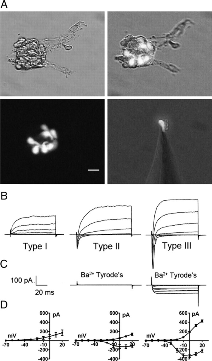
Figure 2.

Optical and electrophysiological identification of taste cell type. A illustrates photomicrographs of circumvallate taste buds and a single isolated taste cell, isolated from a GAD67-GFP mouse, attached to a patch pipette. GFP-labeled cells are type III taste cells. Similarly, type II taste cells could be identified by GFP fluorescence from TrpM5-GFP mice (data not shown). Scale bar, 20 μm. B illustrates typical current profiles of type I, type II, and type III taste cells in Tyrode's. C, Although both type II and type III taste cells have voltage-gated Na+ currents in response to membrane depolarization, only type III taste cells have voltage-gated currents in Ba2+ Tyrode's (with Na+ and K+ currents blocked). D illustrates the averaged I/V curves for the voltage-gated Na+ (circles) and K+ (squares) currents in type I (n = 10), type II (n = 12), and type III (n = 18) cells in Tyrode's. Voltage was stepped in 10 mV increments from −60 to +20 mV from a holding potential of −70 mV. Error bars represent SEM.
