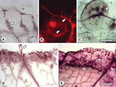
Fig. 2.
Whole mounts showing vascularization associated with early callus development. (A) Venation of a leaf explant cultured for 1 -week and the formation of tracheids at the vein (arrow). (B) A newly formed vein associated with cell proliferation after 2 weeks of culture, initiated at an existing vein and reconnecting to it, forming a circular pattern (arrows). Imaged by fluorescence microscopy. (C) Veins forming in a callus island (arrow) after 2 weeks of culture. (D) A major vein that has grown through the callus and into the medium (arrow) after 1 week of culture. (E) Veins (arrows) growing and branching in the newly formed callus at the edge of the explant after 1 week of culture. Scale bars: (A–C) = 200 µm; (D, E) = 400 µm.

