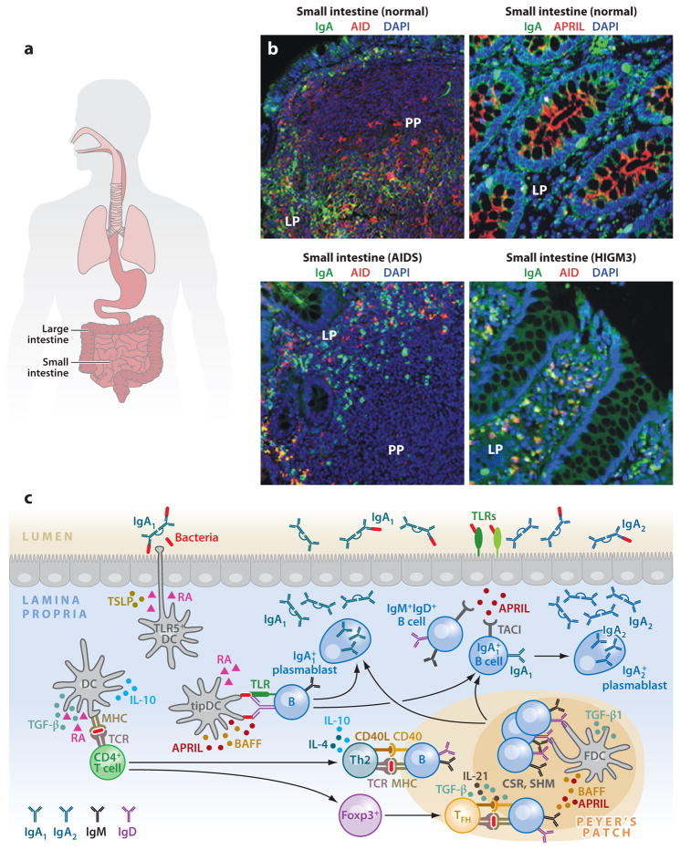
Figure 1.
IgA responses in the intestinal mucosa. (a) Scheme of human MALT, including intestinal mucosa. (b) Immunofluorescence analysis of gut mucosa from healthy, HIGM3, and AIDS donors stained for IgA ( green), AID or APRIL (red ), and nuclei (DAPI staining, blue). Left panels show Peyer’s patches (PPs) and lamina propria (LP); right panels show only LP. Original magnification, ×20. (c) Scheme of mucosal IgA responses. Antigen-sampling DCs receive conditioning signals from TLR-activated intestinal epithelial cells (IECs) via thymic stromal lymphopoietin (TSLP) and retinoic acid (RA). Various DC subsets releasing TGF-β, IL-10, RA, and nitric oxide initiate IgA responses in PPs by inducing Th2, Treg, and Treg-derived T follicular helper (TFH) cells that activate follicular B cells via CD40L, TGF-β, IL-4, IL-10, and IL-21. In addition, DCs activate some B cells in the LP via BAFF, APRIL, and RA. These molecules are also used by TLR-activated IECs to induce local IgA production, including sequential switching from IgA1 to IgA2, as well as plasma cell differentiation and survival. (Additional abbreviations used in figure: AID, activation-induced cytidine deaminase; APRIL, a proliferation-inducing ligand; BAFF, B cell–activating factor; CSR, class switch recombination; DC, dendritic cell; HIGM3, hyper-IgM 3; MALT, mucosa-associated lymphoid tissue; SHM, somatic hypermutation; TGF-β, transforming growth factor-β; TLR, Toll-like receptor.)

