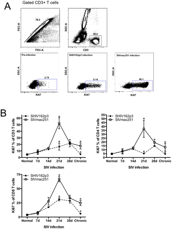
Figure 3. T-cell proliferation during acute SIV infection.
A) Representative gating strategy showing Ki-67 expression on CD3+ T cells prior to and 21 days after infection. B) Longitudinal analysis of early T-cell proliferation in blood from SHIVsf162p3 (filled circles) and SIVmac251-infected RMs (open squares). Means±SEM are shown. Significant differences (P<0.05) between groups are indicated by asterisks.

