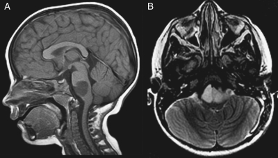
Fig. 2.
One long-term survivor had slightly atypical imaging features for a malignant brainstem glioma, with tumor involvement localized to the pontomedullary junction. Based upon tumor biopsy, the lesion was histologically demonstrated to be an anaplastic astrocytoma. Sagittal T1-weighted (A) and axial T2-weighted (B) MR images demonstrate a T1-hypointense and T2-hyperintense mass at the pontomedullary junction.

