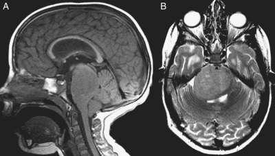
Fig. 3.
MR imaging of one of the 2 long-term survivors with typical imaging features of a diffuse intrinsic brainstem glioma. Sagittal T1-weighted (A) and axial T2-weighted (B) MR images demonstrate a T1-hypointense and T2-hyperintense mass in the pons surrounding the basilar artery with posterior mass effect on the fourth ventricle, moderate hydrocephalus, and tonsillar herniation.

