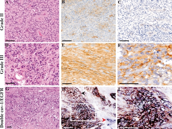
Fig. 1.
Cav-1 and EGFR expression in ependymomas according to tumor grade. One case of Grade II ependymomas (A, H&E, 2×0) that proved to be negative for cav-1 staining (B, ×20). As for the majority of Grade II ependymomas studied, immunohistochemistry for EGFR was negative as well (C, ×20). One case of a Grade III ependymoma (D, H&E, ×20) from our series displaying an intense cav-1 membrane staining (E, ×20) and a diffuse EGFR positivity (F, ×40). In this case of a Grade III ependymoma (G, H&E, ×10), in the majority of positive cells, cav-1 (blue staining) and EGFR (brown staining) were colocalized on the cell membrane, resulting in a black staining (H, ×20; I, ×40). The red arrowhead indicates the internal positive control for cav-1 (blood vessel, in blue); the black arrowhead indicates a group of EGFR-positive/cav-1 negative cells (brown staining). An area of cav-1 and EGFR coexpression is shown in the white frame (bar = 100 μm).

