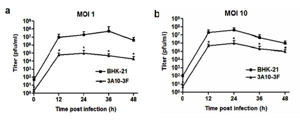
Figure 3.

JEV replication in 3A10-3F (black trianle) and BHK-21 (black square) cells measured by plaque formation assay. Cells were infected with JEV at moi of 1 (a) and 10 (b). The viral titers were determined by plaque formation assay for culture supernatant samples harvested at 0, 12, 24, 36, and 48 hr post-infection. The numbers of virions detected in 3A10-3F cells was greatly reduced. The results displayed were the means of independent experiments performed in duplicate.
