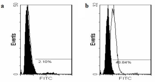
Figure 4.

JEV binding to 3A10-3F and BHK-21 cells measured by flow cytometry. Cells were incubated with JEV for 1 hr and were stained with rabbit anti-JEV antibodies followed by FITC-conjugated goat anti-rabbit IgG. Limited binding of JEV to 3A10-3F cells was shown (2.10%, a), whereas JEV significantly bound BHK-21 cells (48.84%, b).
