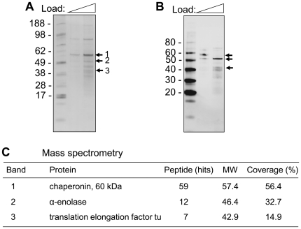
Figure 4. Identification of PLG receptors.
Spore extracts obtained from an acidic buffer were separated on a 4–12% SDS-PAGE. (A) The proteins were visualized by coomassie brilliant blue staining (CBB). (B) Far-Western (Ligand) blot with PLG. The proteins were transferred onto a nitrocellulose membrane, and the membrane was incubated with PLG. PLG-bound proteins were visualized by Western blotting with anti-PLG antibody. (C) Identification of proteins interacting with PLG. Bands (1–3) were excised from the gel and subjected to in-gel digestion with trypsin. LTQ-MS/MS was performed to identify the proteins.

