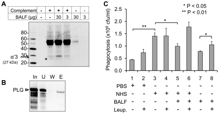
Figure 7. Rabbit BALF decreases spore phagocytosis of macrophages.
(A) Rabbit complements were incubated with BALF (3 and 30 µg) in the PBS for 1 h and subjected to Western blot analysis with anti-C3b antibody. Degradation of C3b was indicated by arrow (27 kDa of α′3 chain). (B) PLG in the BALF interacts with spores. BALF was incubated with spores, eluted by a chaotropic salt and subjected to Western blot analysis with anti-rabbit PLG antibody. (C) BALF decreases NHS-mediated spore phagocytosis. RAW264.7 cells were infected with NHS-treated spores in the presence of BALF and/or leupeptin. Phagocytosed spores were determined by a serial agar plating method.

