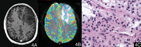
Fig. 4.
A case of oligodendroglioma (WHO grade II). (4A) Postcontrast T1-weighted image showed a nonenhancing lesion in the left frontal lobe. (4B) The color maximal rCBV image overlaid on the T2*-weighted image (transverse gradient-echo dynamic susceptibility-weighted perfusion contrast-enhanced MR image) showed that the maximal rCBV ratio of the tumor was 2.02. (4C) Hematoxylin and eosin, 400×, neoplastic cells show round nuclei and a network of thin capillaries.

