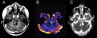
Fig. 1.
Region of interest (ROI) measurements for diffusion and perfusion MR images in brainstem glioma. (A), Axial T1-weighted post-contrast image of a child with brainstem glioma demonstrates expansion of the pons and a region of tumoral enhancement (red arrow) anterior to the fourth ventricle. (B), Cerebral blood volume perfusion map demonstrates a ROI (green circle) chosen on the basis of the highest perfusion value within the tumor. (C), Apparent diffusion coefficient map with the ROI (green circle) placed in an anatomical location closest to that used for perfusion, while avoiding the ventricle.

