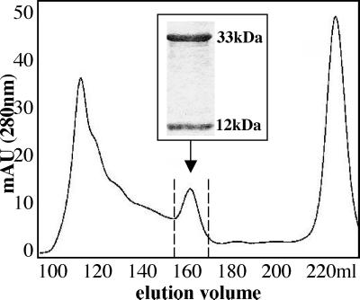
Figure 1.
Purification of refolded CD1d–β2m monomers loaded with α-GalCer. FPLC and UV-light absorption profile at 280 nm wave length of a refolding mix containing CD1d and β2m in the presence of α-GalCer. The arrow indicates the CD1d–β2m monomeric complexes. (Inset) SDS/PAGE analysis of proteins eluted between 155 and 170 ml, which demonstrates the presence of two proteins of 33 and 12 kDa, corresponding to unglycosylated CD1d and β2m, respectively.

