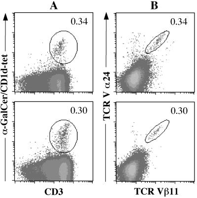
Figure 5.
Staining of intrahepatic lymphocytes in patients with HCV and HBV infection. Samples from patients (P9, top row; P11, bottom row) were stained with CD1d–α-GalCer tetramers and anti-CD3 antibody (A) or anti-Vα24 and -Vβ11 antibodies (B). Percentages of CD1d–α-GalCer tetramer+/CD3+ and Vα24+/Vβ11+ cells are shown.

