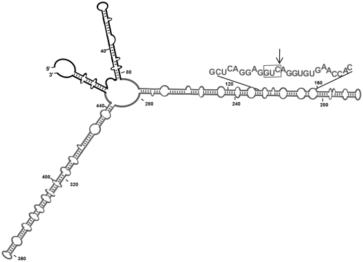
Figure 4.
Schematic representation of the Mfold-predicted secondary structure of the long RNA substrate. Major features of this compact structure have been validated by temperature-gradient gel electrophoresis (35). PSTVd and flanking vector sequences are indicated in grey and black, respectively. The Mfold program (36) and our results obtained with RNase T1 probing (data not shown), indicated that the vector sequences do not disturb the rod-like conformation of the PSTVd(–) RNA. The hammerhead-binding sequence is denoted with capital fonts, with the trinucleotide GUC preceding the cleavage site boxed and the cleavage site marked with an arrow.

