
Figure 8b:
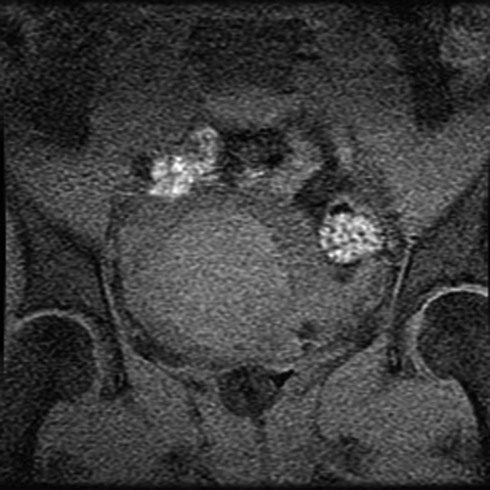
MR-guided FUS of uterine fibroid. (a) Sagittal image shows patient set up and the FUS transducer system on the left. Images (b) before and (c) after intravenous gadolinium administration, acquired immediately after treatment, demonstrate the focal nonperfused fibroid as a result of the ablation.