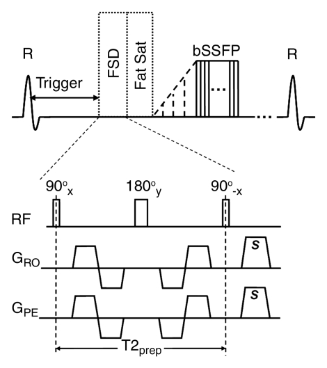
Figure 1a:
Sequence diagrams illustrate (a) dark-artery MR angiography procedure, with FSD-prepared balanced SSFP (bSSFP) triggered at systole and (b) bright-artery MR angiography procedure, with T2-prepared (T2prep) balanced SSFP triggered at diastole. Both FSD preparation and T2 preparation consist of 90°x-180°y-90°x radiofrequency (RF) series and are of the same duration to maintain the same T2 weighting for venous blood and static tissues. FSD gradient pulse applied in readout direction (GRO) and FSD gradient pulse applied in phase-encoding direction (GPE) are simultaneously switched on in FSD preparation but switched off in T2 preparation. Readout direction coincides with main blood flow direction. FSD or T2 preparation is followed by a spectrally selective fat-saturation (Fat Sat) radiofrequency module and 10 preparation radiofrequency pulses with linearly increasing flip angles before the balanced SSFP data acquisition. R = R wave, s = sample.

