
Figure 4.
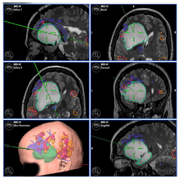
Intra-operative display of pre-and intra-operatively acquired data co-registered to patient and displayed by navigation system 3D viewer. Numbered foreground dots indicate intraoperative cortical stimulation locations. Large green model is segmented tumor. Blue model superior to green is tractography. Orange and red models are functional MRI activations from language tasks in the patient's first and second language. The surgeon can choose a combination of viewpoints which is particularly important to appreciate the three dimensional relations between functional locations and the tumor. Clockwise from top-right: standard orthogonal slices; 3D viewer; axial and sagittal in-line views in the plane of the navigation probe.