
Figure 8.
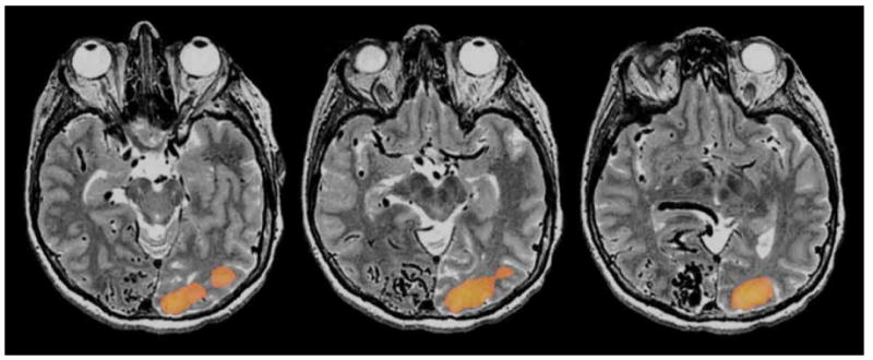
Visual fMRI scans of a patient with a large occipital AVM. A whole filed flashing checkerboard paradigm was delivered. Although the patient had preserved visual perception in the left visual field the patient did not have any visual activation in the right occipital cortex depicted on fMRI