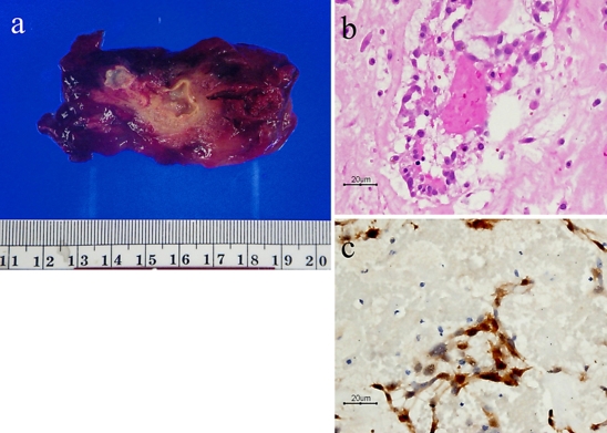
Fig. 2.
A photomicrograph of the 8.0 × 4.7 × 3.7 cm cardiac tumor removed at the operation (a). Microscopically, polygonal and spindle cells were arranged in a single cord or in nests and embedded in the myxoid stroma (HE) (b). The tumor cells show immunoreactivity to calretinin (immunohistochemical stain, anti-calretinin) (c).

