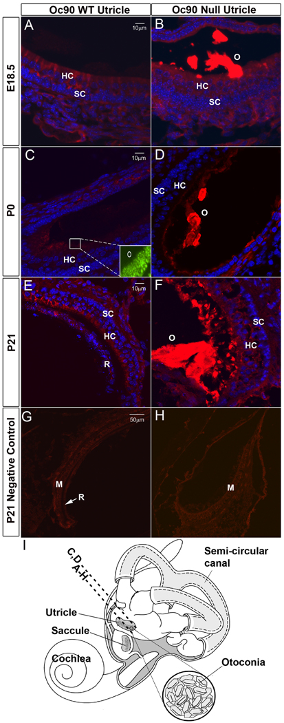
Figure 1.
Deposition of Sc1 in Oc90 wt and null otoconia (the utricle is shown). Immunostaining is shown in (A, B) E18.5, (C, D) P0, (E, F) P21 and (G, H, negative controls at P21 using non-immune serum). In (A, C, E), WT otoconia had extremely faint staining that was slightly visible only under the microscope but not in the photographs, whereas the giant crystals (labeled as “O” in B, D, F) in null tissues were intensely stained. Inset in C shows the presence of normal otoconia in the wt tissue section as detected by Oc90 immunostaining. (I) An illustration of the mouse inner ear. The dashed line labeled with A–H shows the approximate location of the sections except C, D, which are located more periphery as marked by the dashed line above. All epithelial cell types in the utricle and saccule participate in otoconia formation. HC, hair cells; SC, supporting cells; M, macula; R, roof.

