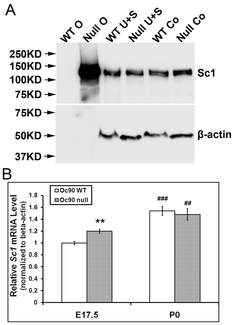
Figure 2.
Otoconial deposition and cellular expression of Sc1 in Oc90 wt and null vestibules. (A) Western blotting of Sc1 in Oc90 wt and null tissues. 6 µg total protein from Oc90 wt otoconia (labeled as “WT O”), 2 µg total protein from null otoconia (“Null O”), and 20 µg total protein from each of the epithelial cellular extracts (U, utricle; S, saccule; Co, cochlea) were loaded. In Oc90 wt otoconia, there was only a faint band after a long exposure; whereas a large amount of Sc1 was detected in Oc90 null otoconia (age P2–3). The same membrane was stripped and re-used for β-actin. β-actin is not present in otoconia, but the equal amount of β-actin detected in each epithelial extract shows that the micro-BCA assay accurately measured the protein content, which was used to control the loading amount. (B) Real-time PCR shows a small but significant increase in Sc1 mRNA in the Oc90 null utricular and saccular epithelia as compared to wt tissues at age E17.5 (**, P<0.01, n=3) but not at P0 (n=3). The relative Sc1 expression levels are higher at P0 than E17.5 in both genotypes (## and ###, P<0.01 and P<0.001, respectively, P0 vs. E17.5 within the same genotype).

