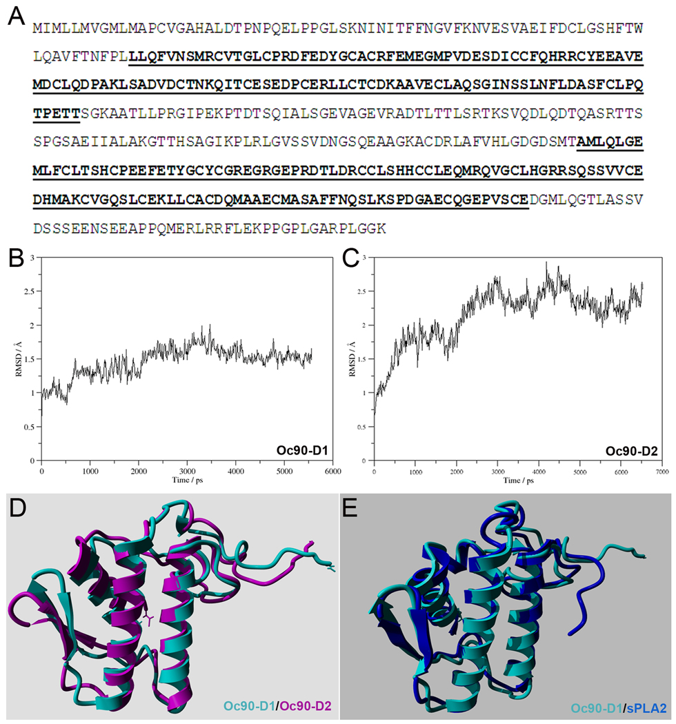
Figure 7.
(A) Amino acid sequence of murine Oc90 (Swiss-Prot id Q9Z0L3). The Pla2l domains are underlined and bolded. (B, C) Backbone RMSD of the first 102 residues as function of simulation time of Oc90-D1 and -D2 (the 1st and 2nd Pla2l domain, respectively). (D) Overlaid time-averaged structures of Oc90-D1 (cyan) and -D2 (purple). (E) Overlaid structures of Oc90-D1 (cyan) and human sPLA2 (blue). The RMSD of the two overlaid structures is 1.517 Å, indicating high structural similarity.

