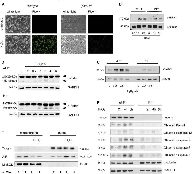
Fig. 3.
Effects of H2O2 on cytosolic Ca2+ and Ca2+ signaling. a Microscopic visualization of intracellular Ca2+ shifts after 5 mM H2O2 (3 h) using the Ca2+ indicator Fluo-4 in wild-type and parp-1 −/− cells. b Western-blot analysis of phosphorylated PERK protein with α-tubulin as loading control. Wild-type (wt P1) and parp-1 knockout MEFs (P1−/−) were treated for the indicated timepoints with 5 mM H2O2. c Western-blot analysis of phosphorylated CaMKII in H2O2 (5 mM) treated wild-type (wt P1) and parp-1 −/− MEFs (P1−/−) at the indicated timepoints. CaMKII is shown as a loading control. d Western-blot analysis of α-fodrin in wild-type (wt P1) and parp-1 knockout (P1−/−) cells. Cells were treated with 5 mM H2O2 for the indicated time points, harvested and subjected to Western-blot analysis. GAPDH is loading control. e Activation of caspases and appearance of cleaved PARP-1 after 5 mM H2O2 is shown in Western-blot analysis of whole cell lysates prepared from wild-type (wt P1) and parp-1 knockout (P1−/−) cells at the indicated timepoints. GAPDH and α-tubulin are shown as loading controls. f Translocation of AIF after H2O2 insult was determined in wild-type cells silenced for the parp-1 gene by RNAi (siRNA 1) and cells silenced with a scrambled control siRNA (siRNA C). After 6 h treatment with 5 mM H2O2, cells were subjected to subcellular fractionation and immunoblotting. Topo-1 and MnSOD protein are also shown

