Figure 2.
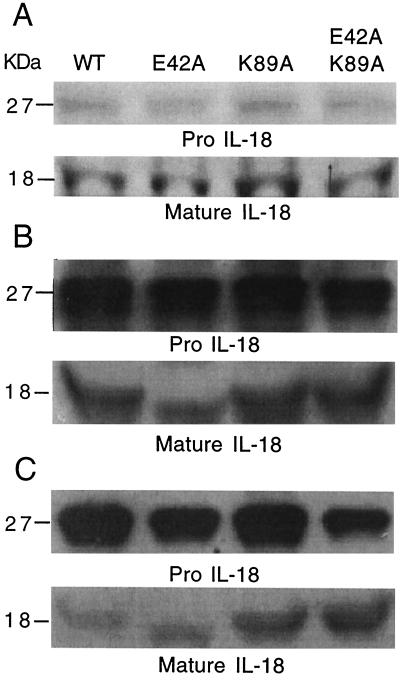
Purified human IL-18 by Coomassie blue staining and Western blot. (A) E. coli expressing the His-6 fusion IL-18 was subjected to 10% SDS/PAGE and stained with Coomassie blue. The numbers on the left indicate molecular masses in kDa. Lanes are identified as WT, E42A, K89A, and E42A/K89A. (Upper) ProIL-18; (Lower) mature IL-18 after factor Xa cleavage. (B) Western blot of preparations shown in A. (Upper) ProIL-18 probed with rabbit anti-human IL-18 (1:500 dilution); (Lower) mature IL-18 probed with the same antibody. (C) Western blot of same preparations shown in A, probed with monoclonal anti-human IL-18 (1:500 dilution of ascitic fluid). (Upper) ProIL-18; (Lower) mature IL-18 probed with the same monoclonal antibody.

