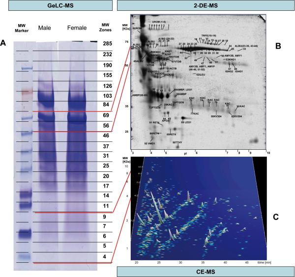
Figure 1. Representative examples of analysis of the human standard urine sample by different proteomics technologies.
A) 1D gel used for fractionation in the GeLC-MS enables separation by mass, and allows for ample material in the subsequent LC-MS/MS analysis of the respective tryptic fragments. The respective molecular weight zones that were excised are indicated on the right. B) 2DE-MS of the female sample, due to its high resolution, enables separation of isoforms/different post-translational modifications. At the same time, representation of the entire gel in the subsequent MS/MS analysis, as in the 1DE, is generally not preformed, due to the size of the gel and the consequently high number of LC-MS/MS analyses required. C) CE-MS (shown is the female sample) enables analysis of the lower molecular weight portion of the urinary proteome that cannot be addressed adequately by SDS-gel based approaches. However, the evidently very high resolution can only be utilized in the analysis of molecules at lower mass, in general < 15 kDa.

