
Fig 4.
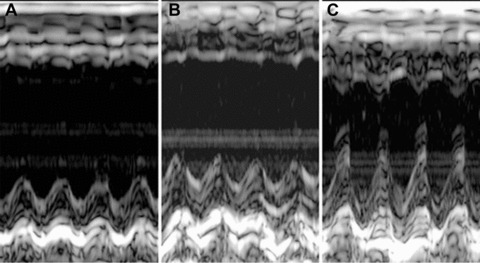
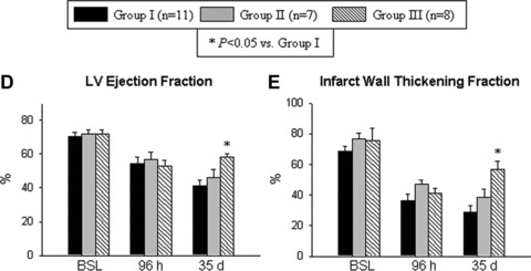
Impact of cell therapy on LV function. (A)–(C) Representative M-mode images at 35 days after coronary occlusion/reperfusion from mice that were given vehicle (group I) (A), expanded and untreated VSEL-SCs (group II) (B) and expanded pre-incubated VSEL-SCs (group III) (C). Compared with the hearts in groups I and II, the heart in group III exhibited a smaller LV cavity, a thicker infarct wall and improved motion of the infarct wall. (D–E) Quantitative echocardiographic analysis revealed improvement in LV functional parameters following transplantation of pre-incubated VSEL-SCs at 35 days after MI. Data are means ± S.E.M. n= 7–11 mice/group. *P<0.05 versus group I at 35 days.