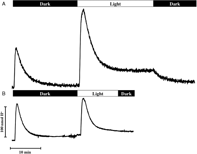
Fig. 2.

Oxygen- and light-induced proton translocation in washed, pigmented (A, OD436 = 9.6) and unpigmented (B, OD436 = 5.7) cell suspensions (see Supporting information S2 for more detail) of Dinoroseobacter shibae incubated under anoxic conditions (see Supporting information S4 for more detail). The first peaks result from the addition of 8 nmol of oxygen in the dark, the second peaks from the addition of 8 nmol of oxygen in the light (average intensity: 420 µE m−2 s−1, see Supporting information S1 for more detail).
