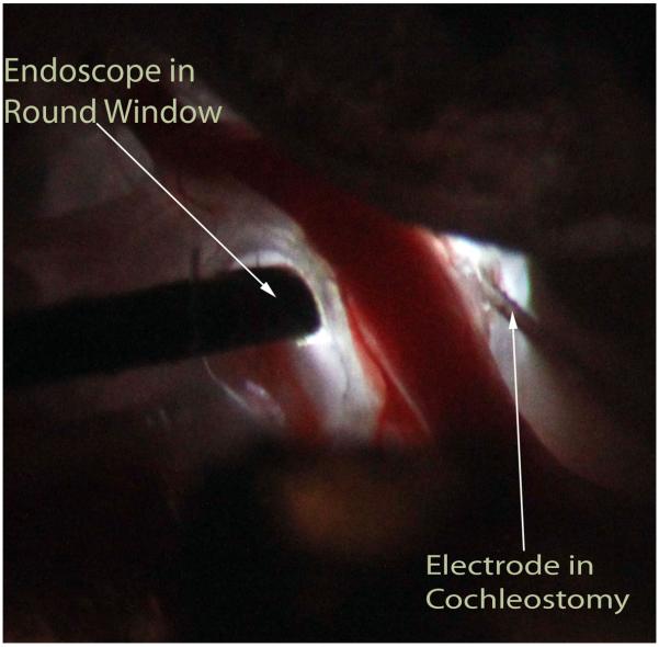
Figure 2.
Right ear of gerbil after posterior exposure of bulla with view of the basal cochlear turns. The light is provided via the endoscope. The endoscope has been placed onto the intact round window membrane while the rigid electrode was inserted through a cochleostomy made in the scala tympani of the basal turn of the cochlea.

